This section will talk about both the organization and major anatomy/physiology of the nervous system. There are three types of neurons: sensory neurons, motor neurons, and interneurons. These are named by their anatomical connections. Sensory neurons are also called afferent neurons, because they pass sensory information from receptors to the nervous system. Motor neurons are also called efferent neurons, because they pass motor commands from the nervous system to muscles to effect an action. Interneurons connect one neuron to another, and are essential to both simple reflex arcs as well as higher order functions such as cognition. A simple reflex arc consists of a sensory neuron, an interneuron, and a motor neuron. As an example, when you step on a nail, receptors in your foot detect the pain and transfer the signal to your nervous system via a sensory neuron. Upon detecting the pain signal, an interneuron in your spinal cord will activate a motor neuron to retract your foot from the source of the pain.
The human nervous system is divided into two sections, the central nervous system and the peripheral nervous system. The central nervous system (CNS) consists of only two parts, the brain and the spinal cord. The 31 pairs of spinal nerves and 12 pairs of cranial nerves, however, are part of the peripheral nervous system (PNS), which is all the nerve tissue and fibers outside the brain and spinal cord. Medical students have all sorts of mnemonics, some NSFW, to help remember the cranial nerves. The olfactory and optic nerves are cranial nerves I and II and are structurally outgrowths of the CNS, but are still considered part of the PNS. The PNS can be further subdivided into the autonomic and somatic nervous systems. The somatic nervous system is straightforward and consists of sensory and motor neurons outside the CNS. The autonomic nervous system (ANS) regulates automatic functions of the body, such as heartbeat, respiration, digestion, and glandular secretions. The ANS can be even further subdivided into the sympathetic and parasympathetic nervous system, which are opposed to each other (antagonistic). The sympathetic nervous system is activated by stress and is responsible for the four F’s: fight, flight, fright, and fuck. The parasympathetic nervous system aims to conserve energy and can be summarized by “rest and digest.” It stimulates peristalsis, increases bile release, and slows heartbeat.
The human brain is perhaps the most complex biological structure in existence. Such an important organ must be properly protected, and the brain is protected by a thick layer of connective tissue called the meninges, which also help anchor the brain to the skull and reabsorb cerebrospinal fluid (CSF). The meninges consist of three layers, which, from internal to external, are the pia mater, arachnoid mater, and dura mater. These layers can be conveniently be remembered with the mnemonic PAD (Pia, Arachnoid, and Dura).
As function follows form, the anatomical divisions of the human brain are similar to its functional divisions. There are three basic subdivisions of the brain: the hindbrain, the midbrain, and the forebrain. Evolutionary, the hindbrain and midbrain developed earlier and are thus responsible for more basic functions of life that aren’t unique to humans. Together, they form the the brainstem, which is the most primitive region of the brain. The midbrain consists of the inferior and superior colliculi is responsible for sensorimotor reflexes. It is located just above the hindbrain. The hindbrain consists of the cerebellum, responsible for precise motor movements, the medulla oblongata, responsible for breathing, digestion, and other vital functions, and the reticular formation, responsible for arousal and alertness, among others. It is located where the brain meets the spinal cord. The forebrain, which contains the neocortex among others, is responsible for emotion, memory, and other complex cognitive and behavioral processes.
While not a focus of the MCAT, it is necessary to have a basic knowledge of the developmental brain. In prenatal life, the brain develops from the neural tube, which is composed of three swellings the correspond to the hindbrain(rhombencephalon), midbrain(mesencephalon), and forebrain(prosencephalon). The hindbrain and forebrain later divide into two swellings each, for a total of 5 swellings. The hindbrain end of the neural tube is connected with the developmental spinal cord.
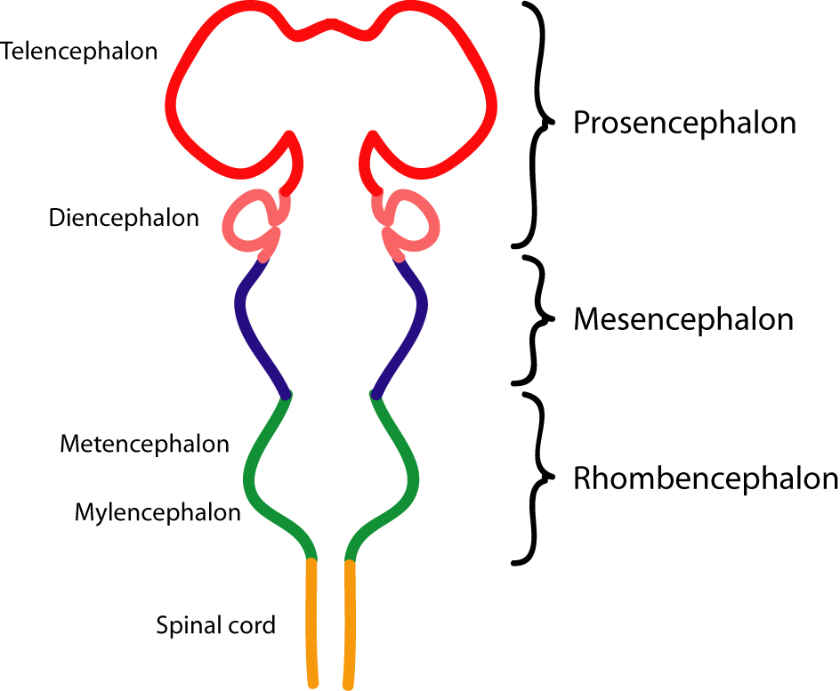
The forebrain is the largest portion of the brain by weight and volume. The thalamus, hypothalamus, posterior pituitary, and pineal gland are derivatives of the diencephalon and the cerebral cortex, basal ganglia, and limbic system are derivatives of the telencephalon. The thalamus is an important sensory relay station in the brain that sorts and transmits incoming sensory information to the appropriate regions of the brain. The hypothalamus is located below the thalamus and is subdivided into the lateral hypothalamus, ventromedial hypothalamus, and anterior hypothalamus. Together, they serve important homeostatic functions by serving to regulate the ANS and endocrine system (The hypothalamus is the primary regulator of the ANS). It also serves key roles in emotional experiences during high arousal states, aggressive behavior, and sexual behavior. The lateral hypothalamus (LH) is referred to as the hunger center that can detect when the body needs more food and fluids and can trigger hunger or thirst. When LH is destroyed in lab mice, they refuse to eat and drink and starve to death. The ventromedial hypothalamus (VMH) is the “satiety center” and provides signals to stop eating. Lesions here lead to obesity. The anterior hypothalamus controls sexual behavior. When stimulated, lab animals will mount just about anything and when lesioned leads to permanent inhibition of sexual activity. It also regulates sleep and body temperature. The pineal gland secretes melatonin which regulates circadian rhythms. The posterior pituitary is the release site for antidiuretic hormone (ADH) which helps the body retain water and oxytocin, which is important for bonding behavior. The basal ganglia coordinate and initiate muscle movement. Parkinson’s disease is associated with destruction of portions of the basal ganglia and is characterized by jerky movements and uncontrolled resting tremors. The extrapyramidal system gathers information about body position (proprioception).
The limbic system is a loop of interconnected structures in the center of the primarily associated with emotion and memory. It includes the septal nuclei, amygdala, and hippocampus. The septal nuclei are one of the primary pleasure centers in the brain and is associated with addictive behavior. The amygdala is associated with defensive and aggressive behaviors, including fear and rage. The hippocampus plays a vital role in learning and memory, helping consolidate information to form long-term memories. Damage to the hippocampus results in anterograde amnesia, or the inability to form new long-term memories. Retrograde amnesia is the loss of previously formed memories. The hippocampus communicates with other portions of the limbic system through the fornix.
The outer surface of the brain is known as the cerebral cortex or neocortex. It has a smooth surface with multiple folds called sulci to maximize surface area. It can be divided into the left and right hemispheres. The major neocortex regions are the frontal lobe, temporal lobe, parietal lobe, and occipital lobe. The frontal lobe is comprised of the prefrontal cortex and the motor cortex. The prefrontal cortex (PFC) is responsible for executive control, which is the regulation of operations of other brain regions. It is associated with perception, memory, emotion, impulse control, and long term planning. Damage to PFC can result in impulsiveness and depression or apathy. This is a good point to highlight the differences between an association area and a projection area. An association area is an area that integrates input from different brain regions while a projection area perform more rudimentary or simple perceptual and motor tasks. The prefrontal cortex is an example of an association area. The visual cortex or motor cortex are examples of projection areas. The primary motor cortex (M1) is located on the precentral gyrus and is responsible for initiating voluntary motor movements. M1 is mapped systematically with parts of the body to which it’s connected, visualized through the motor homunculus, shown below. Certain muscles that require finer motor control occupy more representative space on the homunculus. Broca’s Area is also located in the frontal cortex, and is important for speech production. It is usually unilateral, that is, it is usually only found in one hemisphere. For most people, this is the dominant hemisphere. The left hemisphere is dominant in most people, regardless of handedness.

The parietal cortex is located posterior of the frontal cortex. Among other regions, it contains the somatosensory cortex, which is located on the postcentral gyrus. The somatosensory cortex is similar to the motor cortex and has its own homunculus, shown below. This is also a projection area and is responsible for receiving all incomming sensory signals for touch, pressure, temperature, and pain. The central region of the parietal cortex is associated with spatial processing and manipulation.

The occipital lobe contains the visual cortex, which is a well-understood brain region that receives sensory input from the eyes and processes that information. It is discussed in detail later on, since vision is an important topic. The temporal lobe contains the auditory cortex and Wernicke’s area. The auditory cortex is the primary site of most sound processing and receives input from the inner ears. Wernicke’s area is assocaited with language comprehension. The temporal lobe also has functions in memory processing, emotion, and language.
It is important to appreciate that no lobe or area exists on its own. Only highly interconnected lobes can function properly. In cases where one side of the brain communicates with the other, we call this contralateral communication. For example, motor neurons in the left hemisphere activated muscles in the right side of the body. When cerebral hemispheres communicate with the same side of the body, we call this ipsilateral communication. We also distinguish between dominant and nondominant hemispheres. The dominant hemisphere is typically defined as the one that is more heavily stimulated during language reception and processing. Historically, hand dominance was used as a proxy for hemisphereic domminance, where right-handed individuals were assumed to be left-hemisphere dominant and vice versa. However, only 95% of right-handed individuals are left brain dominant and only 18% of left-handed individuals are right hemisphere dominant. Thus, the dominant hemisphere is usually the left, regardless of handedness. The dominant hemisphere is primarily analytic in function, involved with language, logic, math skills, language production (Broca’s area), and language comprehension (Wernicke’s area). The nondominant hemisphere is associated with intuition, creativity, and spatial processing.